Matrices SOURIS en Acier inox-Dissection cérébrale, coupe coronale ou sagittale-0,5 ou 1 mm
Référence : ND
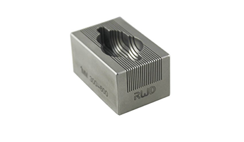
Les matrices sont usinées et traitées pour sectionner des cerveaux de souris avec des intervalles de coupe dans le plan Coronal ou sagittal de 0,5 mm ( 500 microns) ou 1 mm d’épaisseur selon la référence choisie.
Choisissez votre modèle parmi les configurations du menu déroulant :
Ces matrices sont usinées et traitées pour sectionner des cerveaux de souris avec des intervalles de coupes dans le plan coronal ou sagittal. L’épaisseur des tranches obtenu sera de 0,5 mm ( 500 microns) ou 1 mm d’épaisseur selon la référence choisie.
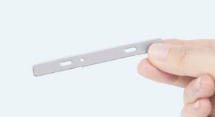
Ce dispositif aide les chercheurs à disséquer des régions spécifiques du cerveau des sujets expérimentaux comme les souris.
Avec ses dimensions anatomiquement proportionnées et ses canaux de tranche, ces matrices sont d’une grande aide pour le chercheur qui souhaite découper des sections régulières dans le plan coronal ou sagittal. Cela permet en outre une fixation précise au froid avant une éventuelle coupe au microtome ou une biopsie pour analyse biochimique.
Par exemple pour la détermination des concentrations de neurotransmetteurs et de métabolites. Des zones cérébrales individuelles peuvent être disséquées ou micro-ponctionnées à partir des tranches formées.
Principales caractéristiques:
– Intervalles de coupe espacés de 500 microns (0,5 mm) ou 1 000 Microns (1 mm).
– Utilisez cette matrice pour sectionner l’organe en tranches coronales ou sagittales avec des épaisseurs par incréments de 0,5 mm ou 1 mm.
– Conçu pour s’adapter aux caractéristiques anatomiques uniques du cerveau de l’espèce sélectionnée, y compris le tissu olfactif/spinal adjacent.
– Inclus un canal de coupe sagittale central permettant de diviser le cerveau en hémisphères gauche et droit.
– A utiliser avec des tissus cérébraux frais ou bien préservés.
– Matrice autoclavable et conçu pour une utilisation sur le long terme.
– Les matrices d’un type donné sont identiques d’un fabrication à l’autre et assurent ainsi des sections reproductibles.
– Livrées prête à l’emploi.
– en acier inoxydable de haute qualité avec polissage en post-traitement pour une grande durabilité.
-Peut être chauffé, refroidit, auto-clavé et utilisé dans une grande variété de conditions comme plongé dans une solution de chlorure de tétrazolium.
Les utilisations comprennent:
– Dissection/blocage du cerveau dans des coupes coronales (ou hémisphères gauche et droit).
– Sectionner le cerveau avant ou après la coloration avec des agents comme le CTT ou l’X-Gal-Stain.
– Isolement répété des régions cérébrales pour des analyses ultérieures ou la préparation de lames
– Préparation des tissus
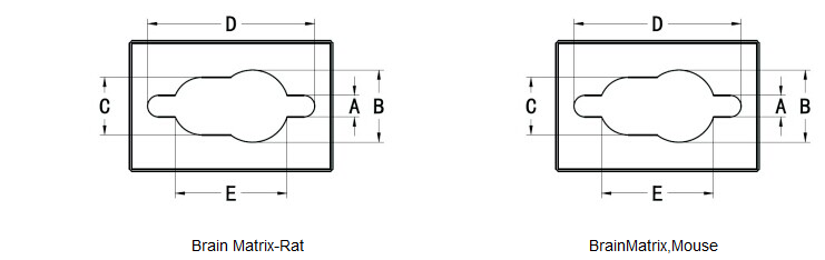
Cat No.
Description de la matrice
A
B
C
D
E
Profondeur
(mm)
(mm)
(mm)
(mm)
(mm)
(mm)
VI-68707
Matrice de cerveau , souris 40-75g, Coronale, 1mm, inox
3.18
11.1
8.73
19.1
12.5
7.4
VI-68708
Matrice de cerveau , souris 40-75g, sagittale, 1mm, inox
3.18
11.1
8.73
19.1
12.5
7.4
VI-68713
Matrice de cerveau , souris 40-75g, Coronale, 0,5 mm, inox
3.18
11.1
8.73
19.1
12.5
7.4
Données techniques
Poids
0.4 kg
Dimensions
3 × 2 × 2 cm
matériaux
Inox polis
section
0,5 mm ou 1 mm selon modèles
précautions
aucune autoclavable peut être monté en température, refroidit, trempé dans des solutions chimiques divers compatibles avec l'inox
The combination of astragalus membranaceus and ligustrazine ameliorates micro-haemorrhage by maintaining blood–brain barrier integrity in cerebrally ischaemic rats
Jun Cai et al. Journal of Ethnopharmacology 2014
Haemorrhagic transformation is an asymptomatic event that frequently occurs after following ischaemic stroke, particularly when pharmaceutical thrombolysis is used. However, the mechanism responsible for haemorrhagic transformation remains unknown, and therapeutics have not been identified. In this study, we administered a combination of astragalus membranaceus and ligustrazine to rats with cerebral ischaemia that had undergone thrombolysis. We analysed the effect of this combination on the attenuation of haemorrhagic transformation and the maintenance of blood–brain barrier integrity.
A longitudinal study of the mechanical properties of injured brain tissue in a mouse model
Yuan Feng et al. Journal of the Mechanical Behavior of Biomedical Materials 2017
Mechanical properties of brain tissue are crucial to understand the mechanism of traumatic brain injury (TBI). Over the past several decades, most of the studies focused on healthy brain tissues, while few of them are about the injured tissues. Therefore, limited knowledge is known about the mechanical properties of the injured brain tissues. In this study, we used an in vivo mouse model with a weight drop device to study injured brain tissues. Around the injury site, mechanical properties of the injured, neighboring, and the corresponding contralateral regions of interest (ROIs) were measured over five temporal points by indentation. Longitudinal and regional comparisons of the mechanical properties revealed that the ROI of the injured tissue had a higher elastic modulus than the contralateral counterpart one-hour post-injury. However, the elastic modulus decreased one-day post-injury and recovered to be close to the contralateral ROI in 7 days. The elastic modulus curves of the injured and the contralateral counterpart ROIs crossed at time points of 12 h and 1 day post-injury, where two significant increases of glial fibrillary acidic protein (GFAP) positive cells were observed. Biological staining results indicated that both the astrocytic responses and the morphological structure could affect the mechanical properties of the injured tissue. The observed longitudinal changes of the mechanical properties at the tissue level and the morphological and biological changes at the cellular level provide insights into understanding the mechanism of TBI. Results are also meaningful for applying emerging in vivo diagnostic tools such as magnetic resonance elastography (MRE) in TBI detection.
SCM-198 protects ischemic brain injury via accelerating the recovery of brain glucose metabolism, ameliorating the damage of neurons and inhibiting the activation of microglia
Jie Xu et al. Int J Clin Exp Med 2017
SCM-198 was chemically synthesized based on the structure of Leonurine and have been found to be effective in prevention of ischemic stroke; However, its therapeutic effect on ischemic stroke was poorly understood. In this study, we used rats to build up permanent middle cerebral artery occlusion (MCAO) model to determine the therapeutic effects and potential mechanism of SCM-198 on ischemic injury. 2,3,5-triphenyltetrazolium chloride (TTC) staining, neurological deficit evaluation, 18 F-FDG PET/CT scanning, transmission electron microscopy (TEM) and immunohistochemical staining were employed in the present study. TTC staining and neurological deficit evaluation results revealed that, in a 2 h window of opportunity, infarct volume and neurologic deficit scores were both decreased after SCM-198 treatment. In the PET/CT scanning study, we found the Ipsilateral-to-Contralateral ratio of all SCM-198 treatment groups were bigger than that of the MCAO group. In the TEM and immunohistochemical staining parts, we found the morphology of neurons was improved and the activation level of microglia was inhibited after SCM-198 treatment. Those results indicated that SCM-198 was a potential therapeutic option for ischemic injury in a 2 h window of opportunity, which may be related with its effects in recovering the brain glucose metabolism, ameliorating the damage of neurons and inhibiting the activation of microglia after ischemic injury happened.
Alzheimer mouse brain tissue measured by time resolved fluorescence spectroscopy using single‐ and multi‐photon excitation of label free native molecules
Bidyut Das et al. journal of biophotonic 2017
Time resolved spectroscopic measurements with single‐photon and multi‐photon excitation of native molecules were performed ex vivo on brain tissues from an Alzheimer’s disease (AD) and a wild type (WT) mouse model using a streak camera. The fluorescence decay times of native NADH and FAD show a longer relaxation time in AD than in WT tissue, suggesting less non‐radiative processes in AD. The longer emission time of AD may be attributed to the coupling of the key native building block molecules to the amyloid‐tau and/or to the caging of the native fluorophores by the deposition of amyloid‐beta or tau plaques and neurofibrillary tangles that affect the local non‐radiative interactions.
Exogenous fractalkine enhances proliferation of endothelial cells, promotes migration of endothelial progenitor cells and improves neurological deficits in a rat model of ischemic stroke
Wenjing Qin et al. Neuroscience Letters 2014
Fractalkine/CX3CL1, also called neurotactin, has been described as an angiogenic agent, and its expression is up-regulated in the penumbra after ischemia. This study was conducted to investigate the neovascular potential of fractalkine on rat models of transient middle cerebral artery occlusion (MCAO). Rats receiving intracerebroventricular injections of fractalkine were found to have improved neurological deficits, reduced cerebral infarct size and increased neuron survival for both doses (100 ng and 1 μg). Fractalkine exerted angiogenic effects that showed dose-dependent higher vascular densities in the peri-infarct area. Furthermore, exogenous fractalkine increased the proliferation of endothelial cells in a dose-dependent manner and enhanced the migration of endothelial progenitor cells at the higher dose (1 μg) in ischemic penumbra. In conclusion, intracerebroventricular administration of fractalkine reduces ischemic damage by promoting neuroprotection and by inducing endothelial cell proliferation and endothelial progenitor cell migration, thereby enhancing neovascularization in the peri-infarct region.
Label-Free Fluorescence Spectroscopy for Detecting Key Biomolecules in Brain Tissue from a Mouse Model of Alzheimer’s Disease
Lingyan Shi et al. Scientific Reports volume 2017
In this study, label-free fluorescence spectroscopy was used for the first time to determine spectral profiles of tryptophan, reduced nicotinamide adenine dinucleotide (NADH), and flavin denine dinucleotide (FAD) in fresh brain samples of a mouse model of Alzheimer’s disease (AD). Our results showed that the emission spectral profile levels of tryptophan and NADH were higher in AD samples than normal samples. The intensity ratio of tryptophan to NADH and the change rate of fluorescence intensity with respect to wavelength also increased in AD brain. These results yield an optical method for detecting early stage of AD by comparing spectral profiles of biomolecules.
Nous utilisons des cookies pour vous garantir la meilleure expérience sur notre site. Si vous continuez à utiliser ce dernier, nous considérerons que vous acceptez l'utilisation des cookies.Ok