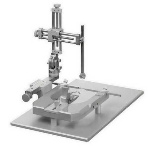
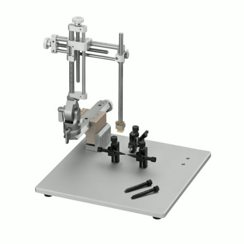
Cadre stéréotaxique portable (design allégé) pour Rat
Référence : ND
Cadre de stéréotaxie compact développé pour la contention sous anesthésie des Rats. Il combine toutes les caractéristiques attendues pour une table stéréotaxique mais avec une faible emprunte de paillasse.
Choisissez votre modèle parmi les configurations du menu déroulant :
Harmony of T cell profile in brain, nasal, spleen, and cervical lymph nodes tissues in Alzheimer’s: A systemic disease with local manifestations addremove
International Immunopharmacology
https://doi.org/10.1016/j.intimp.2020.107306
NafisehPakravan et al.
14 December 2020
Neural repair by NT3-chitosan via enhancement of endogenous neurogenesis after adult focal aspiration brain injury addremove
Determination of dexmedetomidine using high performance liquid chromatography coupled with tandem mass spectrometric (HPLC-MS/MS) assay combined with microdialysis technique: Application to a pharmacokinetic study addremove
Cadre de stéréotaxie compact et allégé, développé pour la contention sous anesthésie des Rats. Il combine toutes les caractéristiques attendues pour une table stéréotaxique de paillasse. Un micromanipulateur tridimensionnel à lecture par verniers ( 100 microns) ou numérique ( 10 microns). Livré en standard avec un ou deux micro-manipulateurs.
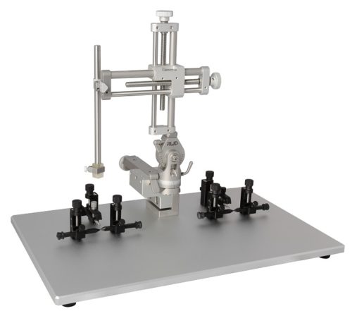
Barres d’oreille, adaptateur de gueule et porte accessoire sont à choisir séparément:
injection stéréotaxique rat, de nouvelles formulations médicamenteuses, molécules inhibiteurs et cultures cellulaire
Mise en place de Microdialyse cérébrale
placement pour Stimulation nerveuse et enregistrement du signal EEG
Optogénétique et fiber photométrie
Imagerie cérébrale
Modélisation des lésions cérébrales et de la moelle épinière
——–
Avec les accessoires adéquats, ce cadre peut être adapté à plusieurs espèces, souris& rats notamment.
La gravure laser des échelles est travaillée de manière à réduire la fatigue visuelle, des icônes dé-trompeuses permettent de prendre en main le positionnement rapidement.
Ajustement avant-arrière de la barre de gueule de 30 mm en hauteur de 43,5 mm avec vernier de 1 mm de résol.
Remarque:
Cette table ne dispose pas de support en U rendant l’ensemble plus léger et maniable, la rigidité est assurée par une double fixation par vis M6.
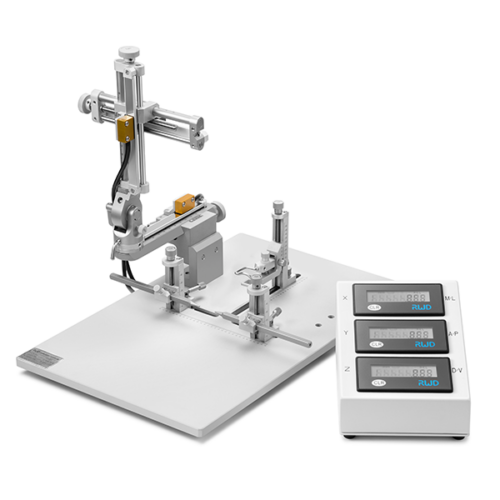
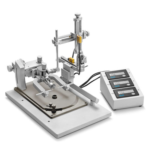
Ce cadre peut être équipé d’un micro-positionneur à verniers digitaux pour en augmenter la résolution:
cadre rats portable avec micromanipulateur digital
Données techniques
Poids
8 kg
Dimensions
40 × 25.5 × 30 cm
Hauteur des barres d'oreilles
35.5 mm
Résolution positionnement barres d'oreilles
0.1 mm
Plage ajustement barres d'oreilles
35 mm
Plage ajustement barre de gueule
Dorsale/ Ventrale 30 mm (+10mm~-20mm) avec une précision de 0.1mm.
GABAergic ventrolateral pre‑optic nucleus neurons are involved in the mediation of the anesthetic hypnosis induced by propofol
Jie Yuan et al. Molecular Medicine Reports 2017
Intravenous anesthetics have been used clinically to induce unconsciousness for seventeen decades, however the mechanism of anesthetic‑induced unconsciousness remains to be fully elucidated. It has previously been demonstrated that anesthetics exert sedative effects by acting on endogenous sleep‑arousal circuits. However, few studies focus on the ventrolateral pre‑optic (VLPO) to locus coeruleus (LC) sleep‑arousal pathway. The present study aimed to investigate if VLPO is involved in unconsciousness induced by propofol. The present study additionally investigated if the inhibitory effect of propofol on LC neurons was mediated by activating VLPO neurons. Microinjection, target lesion and extracellular single‑unit recordings were used to study the role of the VLPO‑LC pathway in propofol anesthesia. The results demonstrated that GABAA agonist (THIP) or GABAA antagonist (gabazine) microinjections into VLPO altered the time of loss of righting reflex and the time of recovery of righting reflex. Furthermore, propofol suppressed the spontaneous firing activity of LC noradrenergic neurons. There was no significant difference observed in firing activity between VLPO sham lesion and VLPO lesion rats. The findings indicate that VLPO neurons are important in propofol‑induced unconsciousness, however are unlikely to contribute to the inhibitory effect of propofol on LC spontaneous firing activity.
Neural repair by NT3-chitosan via enhancement of endogenous neurogenesis after adult focal aspiration brain injury
PengHao et al. Biomaterials 2017
The latent regenerative potential of endogenous neural stem/progenitor cells (NSCs) in the adult mammalian brain has been postulated as a likely source for neural repair. However, the inflammatory and inhibitory microenvironment after traumatic brain injury (TBI) prohibits NSCs from generating new functional neurons to restore brain function. Here we report a biodegradable material, chitosan, which, when loaded with neurotrophin-3 (NT3) and injected into the lesion site after TBI, effectively engaged endogenous NSCs to proliferate and migrate to the injury area. NSCs differentiate and mature into functional neurons, forming nascent neural networks that further integrate into existing neural circuits to restore brain function. Three main actions of NT3-chitosan, i.e., pro-neurogenesis, anti-inflammation, and pro-revascularization, elicit significant regeneration after TBI. Our study suggests that through creating an optimal microenvironment, endogenous NSCs are capable of executing neural repair, thus widening the therapeutic strategies to treat TBI and perhaps stroke or other neurological conditions.
Nous utilisons des cookies pour vous garantir la meilleure expérience sur notre site. Si vous continuez à utiliser ce dernier, nous considérerons que vous acceptez l'utilisation des cookies.Ok