Publications
Traditional Chinese Medicine Huannao Yicong Decoction Extract Decreases Tau Hyperphosphorylation in the Brain of Alzheimer’s Disease Model Rats Induced by Aβ1–42
Yu Cao Evidence-Based Complementary and Alternative Medicine 2016
Huannao Yicong Decoction has been shown to improve the learning and memory capabilities of Alzheimer’s disease (AD) subjects. However, the underlying mechanism remains to be determined. Methods. Sixty Sprague-Dawley rats were divided equally and randomly into five different groups including control, positive control, and HYD granules of low dose, medium dose, and high dose by daily gavage. The sham-treated rats were also given the same volume of sterile water by gavage. Twelve SD rats were treated with the same amount of physiological saline. Twelve weeks later, learning and memory capabilities, Aβ content of the right brain and the expression of glycogen synthase kinase-3β (GSK-3β), total tau protein kinase (TTBK1), and cyclin-dependent kinase-5 (CDK-5) were tested. Results. Our results showed that high dose HYD treatment significantly improved the learning and memory capability of the AD rats and decreased the expression of TTBK1, GSK-3β, and CDK-5 in the hippocampal CA1 region. Conclusions. HYD treatment for 12 weeks significantly improved spatial learning and memory and effectively inhibited Aβ deposition, likely via reducing tau protein kinase expression and thus tau hyperphosphorylation and inflammatory injury. Taken together, these results suggest that HYD could be an effective treatment for AD.
Glioma Growth Inhibition In Vitro and In Vivo by Single Chain Variable Fragments of the Transferrin Receptor Conjugated to Survivin Small Interfering RNA
F Wang et al. Journal of International Medical Research 2011
Inhibition of survivin by small interfering RNA (siRNA) can delay glioma growth. To enhance the effect of survivin-siRNA on intercranial glioma, a conjugate of siRNA-survivin and single-chain variable fragment (scFv) of the transferrin receptor (TfR) was constructed and its effects assessed in vitro and in vivo. Survivin-siRNA and the survivin-siRNA/scFv-TfR conjugate were constructed and used successfully to inhibit glioma U87 cell proliferation and enhance apoptosis in vitro. The molecular constructs were administered to an established U87 orthotopic mouse model. Western blot analysis and immunohistochemistry of the glioma tissues showed that survivin protein levels were strongly suppressed by the survivin-siRNA/scFv-TfR conjugate in vivo. Furthermore, survivin-siRNA/scFv-TfR prolonged the survival time of mice more than survivin-siRNA. In conclusion, the survivin-siRNA/scFv-TfR conjugate efficiently enhanced the effects of survivin-siRNA on glioma suppression in vivo, confirming the applicability of antibody-targeted molecular therapies for treating human brain tumours.
Protective Effect of Klotho against Ischemic Brain Injury Is Associated with Inhibition of RIG-I/NF-κB Signaling
Hong-Jing Zhou et al Front. Pharmacol 2018
Aging is the greatest independent risk factor for the occurrence of stroke and poor outcomes, at least partially through progressive increases in oxidative stress and inflammation with advanced age. Klotho is an antiaging gene, the expression of which declines with age. Klotho may protect against neuronal oxidative damage that is induced by glutamate. The present study investigated the effects of Klotho overexpression and knockdown by an intracerebroventricular injection of a lentiviral vector that encoded murine Klotho (LV-KL) or rat Klotho short-hairpin RNA (LV-KL shRNA) on cerebral ischemia injury and the underlying anti-neuroinflammatory mechanism. The overexpression of Klotho induced by LV-KL significantly improved neurobehavioral deficits and increased the number of live neurons in the hippocampal CA1 and caudate putamen subregions 72 h after cerebral hypoperfusion that was induced by transient bilateral common carotid artery occlusion (2VO) in mice. The overexpression of Klotho significantly decreased the immunoreactivity of glial fibrillary acidic protein and ionized calcium binding adaptor molecule-1, the expression of retinoic-acid-inducible gene-I, the nuclear translocation of nuclear factor-κB, and the production of proinflammatory cytokines (tumor necrosis factor α and interleukin-6) in 2VO mice. The knockdown of Klotho mediated by LV-KL shRNA in the brain exacerbated neurological dysfunction and cerebral infarct after 22 h of reperfusion following 2 h middle cerebral artery occlusion in rats. These findings suggest that Klotho itself or enhancers of Klotho may compensate for its aging-related decline, thus providing a promising therapeutic approach for acute ischemic stroke during advanced age.
A reinforced composite structure composed of polydiacetylene assemblies deposited on polystyrene microspheres and its application to H5N1 virus detection
Wenjie Dong et al. Int J Nanomedicine 2013
In this study, we immobilized polydiacetylene vesicles (PDAVs) onto the surface of polystyrene (PS) microspheres (1 μm in diameter) by using both electrical charge and conjugated forces to form a reinforced composite structure. These reinforced complexes could be easily washed, separated by centrifugation, and resuspended by gentle agitation. After passing through a narrow 200 μm-diameter channel, the composite structures maintained their original shape, demonstrating their resilience and potential for use in microfluidic technologies. The number of PDAVs in the composite structure could be mediated by changing the extent of layer deposition, which affected the sensitivity of detection. It showed that PDAVs did not change their blue color after addition of detecting probes such as anti-H5N1, which was of great importance in the fabrication and modification of stable color-changeable biosensors based on PDAVs. By conjugating anti-H5N1 antibodies to the PS@PDAV via N -hydroxysuccinimide and 1-ethyl3-(3-dimethylaminopropyl) carbodiimide chemistry, a stable blue complex, anti-H5N1 microsphere (PS@PDAV-anti-H5N1) was formed. A target antigen of H5N1 (HAQ [H5N1 strain A/ environment/Qinghai/1/2008{H5N1} in clade 0]) was detected by PS@PDAV-anti-H5N1. At an optimal PDAV deposition level of three layers, the limit of detection was determined to be approximately 3 0 ng/mL of HAQ by using optical spectrum measurement and visual inspection, meeting the needs of fast and simple color-changeable detection. However, a much lower limitation of detection (1 ng/mL) was able to be obtained using laser-scanning confocal microscopy, which could be compared with the results obtained with other sophisticated equipment.
An immunoassay in which magnetic beads act both as collectors and sensitive amplifiers for detecting antigens in a microfluidic chip (MFC)–quartz crystal microbalance (QCM) system
JianhuaHan et al.Colloids and Surfaces A: Physicochemical and Engineering Aspects 2011
An immunoassay based on the enrichment of antibodies by magnetic beads (MB) and their subsequent detection of antigens in a microfluidic chip (MFC)–quartz crystal microbalance (QCM) was developed and investigated. The MB conjugated to goat anti-human-immunoglobulin G (MB-anti-h-IgG) were concentrated within microchannels by the application of a magnetic field, which also functioned as a separation switch outside the MFC. The conjugation of the MB and the anti-h-IgG antibody in the MFC was completed in 1 min. Removal of the magnetic field allowed the resulting tributary buffer solution to flush the MB-anti-h-IgG out of the MFC and onto a QCM chip. The combination of the MB-anti-h-IgG and the QCM chip amplified the detection sensitivity of antigen(s) to 10−1 ng/ml. By controlling the enrichment velocity and sample injection mode in the MFC, a linear response ranging from 10−1 to 103 ng/ml was obtained. This technique establishes a simple, rapid MFC method for the amplification of a QCM-based immunoassay.
A novel three-dimensional microfluidic platform for on chip multicellular tumor spheroid formation and culture
Duanping Sunet al Microfluidics and Nanofluidics 2014
The formation of three-dimensional (3D) multicellular cell spheroids such as microspheres and embryoid bodies has recently gained much attention as a useful cell culture technique, but few studies have investigated the suitability of glass for spheroids formation and culture. In this work, we present a novel three-dimensional microfluidic device made of poly(dimethylsiloxane) (PDMS) and glass for the easy and rapid synthesis and culture of tumor spheroid. The cell culture unit is composed of an array of microwells on the bottom of a glass plate, bigger microwells and elastomeric microchannels on the top of a PDMS plate. Cell suspension can be easily introduced into the cell culture unit and exchange with the external liquid environment by the microfluidic channels. A single tumor spheroid can be formed and cultured in each glass cell culture chamber, the surface of which was modified with poly(vinyl alcohol) to render it to be resistant to cell adhesion. As the cell culture medium could be replaced, spheroids of the human breast cancer (MCF-7) cells were cultured on the chip for 3 days, reaching the diameters of about 150 μm. Furthermore, the MCF-7 cells were successfully cultured on the chip in 2D and 3D culture modes. Results have shown that glass is well suitable for multicellular tumor spheroids culture. The established platform provides a convenient and rapid method for tumor spheroid culture, which is also adaptable for anticancer drug screening and fundamental biomedical research in cell biology.
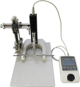
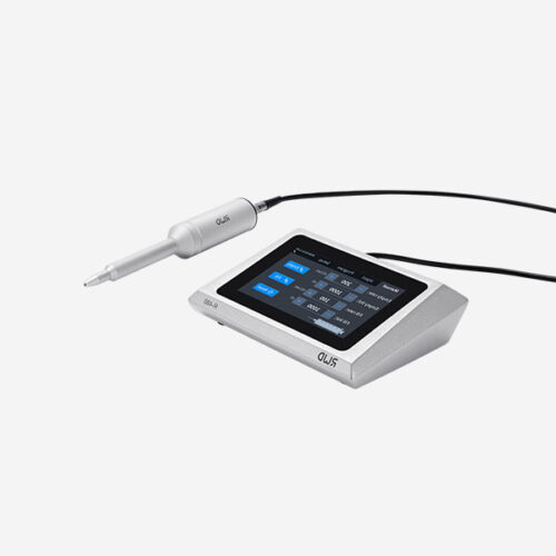
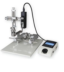 micromanipulateur d’un cadre de stéréotaxie.

